Profile


Dr Pankaj Goel, is currently working as a Director and Head of the Cardio-thoracic and Vascular (Heart, Lung and Vascular Surgery) unit at the Ivy Hospital, Amritsar.

After completing his MCh in cardio-thoracic and vascular surgery from GB Pant Hospital, Delhi University in 1998, Dr Goel worked at Madras Medical Mission, Chennai for three years. Here he obtained training in complex paediatric cases. Thereafter he went to Australia (Royal Perth Hospital) for further training and experience.

Dr Goel joined the Fortis Escorts Hospital, Amritsar in 2003. Since 2008 , in his capacity as HOD at the same hospital he has done pioneering work and established cardio-thoracic and vascular surgery in the city.

The Goel's unit now routinely performs all types of cardiac, thoracic and vascular surgeries with results comparable to the best centres in the world. Dr Goel is responsible for many firsts in the region.

Dr Goel has several research papers published in indexed journals. He has authored a book on cardiac surgery. He has an original technique for harvesting saphenous vein to his credit.

In 2009, Dr Goel was elected member of the prestigious Society of Thoracic Surgeons , USA. He is also a member of the Indian Association of Cardio-thoracic surgery and CTS Net.

At Ivy Hospital, Dr Goel routinely performs all types of Cardiac, thoracic (including thoracoscopic) and vascular procedures.


Tuesday 11 December 2012

Case of the Month- Post MI Posterior LV Aneurysm- Endoventricular patch Repair.

Case of the Month- Post MI Posterior LV Aneurysm- Endoventricular patch Repair
 
A sixty year old diabetic male presented to us with a three week history of chest discomfort accompanied by sweating, restlessness and uneasiness.
He was evaluated elsewhere for coronary artery disease but the echo was found to be unremarkable. His chest X- ray revealed left pleural effusion, and he was empirically started on ATT. The patient did not improve and came to us for review/second opinion.

We conducted the following tests:

ECG- t wave inverted infr. Leads                                                                                              


Echo-
 Posterior LV aneurysm with pericardial effusion.

 Coronary Angio-                Occluded RCA.                      
 Proximal LAD 70-80%.


In view of the echo findings and the symptoms, a diagnosis of post MI, aneurysm with contained rupture of the posterior LV wall was made.

A coronary angiography was performed, which revealed significant lesions of LAD and complete occlusion of RCA.

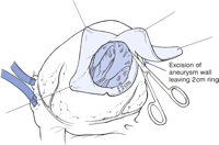
The patient was taken up for emergency surgery. Per-operatively there were dense adhesions in the pericardium. A large 6cmX3cm true aneurysm of the posterior LV wall was found. The patient underwent CABGX2 with LIMA to LAD and SVG to PDA. The aneurysm was excised and defect in the left ventricle repaired with a Dacron patch. (Endoventricular patch). The patients lt pleural fluid was sent for examination and was negative for tuberculosis.


The patient made an uneventful recovery. The post- operative echo shows-  normal lv function, minimal pericardial effusion (post surgery).

Comment-
LV aneurysms complicate 10% of MI’s. Most of them have a chronic course and a good prognosis on medical management. Surgery is indicated when symptoms appear. The usual symptoms are, - angina, Dyspnoea and ventricular arrhythmias causing syncope. On echo an EDV of more than 120ml/m2 is also a relative indication. Rupture is a life threatening condition requiring urgent surgery.

Indications for Surgery.

Documented expansion/large size
Angina
Congestive heart failure
Arrhythmia
Rupture
Pseudoaneurysm
Congenital aneurysm
Embolism
Documented expansion/large size


Surgery consists of CABG (if there is significant CAD on angiography) along with excision of aneurysm and repair of defect in LV with a Dacron patch. The operative mortality is 4-7% with a 80% five year survival.


Monday 10 December 2012

Punjab's First freestyle Porcine Valve Implant.

Last year we operated on a fifteen year old girl with severe aortic stenosis. She had the rare birth defect of unicuspid aortic valve. Her heart function had come down to 25% and she could barely walk. Her annulus size and ascending aorta were very small. Implanting a regular mechanical valve would have been futile as she would have high residual gradients, also she would have to be on life long anticoagulation.
I therefore implanted a freestyle valve as a complete root replacement technique. This valve is a commercially available pig valve. In a sense it is like transplanting a pig valve in the patient. The advantages are that there are minimal gradients and the heart function recovers promptly. Also the girl need not be on anticoagulation and can have a normal family life. Needless to say the procedure is technically very demanding and time consuming.
At one year follow up the girl is healthy with normal heart function.
The company reps tell me that this is the first time such a valve has been implanted in Punjab!!!


Freestyle Porcine(pig) aortic valve.